For a long time, doctors accepted they had under six hours later a stroke started to keep a patient’s synapses from biting the dust and to save the patient from death or inability.
Be that as it may, from the start of his vocation, Gregory Albers, MD, educator of nervous system science and neurosurgery, scrutinized this standard way of thinking. Throughout the span of thirty years, he and a group of Stanford Medicine specialists tried and culminated another imaging method that would, at last, grow the window of time for securely reestablishing bloodstream in a stroke patient’s mind.
In “Opening stroke’s window,” a story in the most recent issue of Stanford Medicine magazine, Albers’ excursion to assist treat with additional stroking patients spreads out, featuring how only a couple of hours can have a major effect on patients’ lives.
“It was an immense test,” Albers said of his journey to further develop results for stroke patients. “We were managing the No. 1 reason for incapacity around the world, and there was no treatment.”
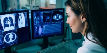
At the point when Albers started his exploration, it took doctors hours to get pictures of a stroke patient’s mind – – and by then the harm had effectively been finished. He and his group found a substitute method for seeing which synapses had been compromised and distinguish assuming any tissue could, in any case, be saved on the off chance that doctors mediated.
As the group balanced their underlying trial of the imaging, the information recommended that with regards to half of the stroke patients could in any case benefit assuming the coagulation obstructing their cerebral supply route was eliminated as long as 12 hours later stroke side effects started. Obviously, there was more work to be finished. Albers and his group proceeded with a progression of preliminaries, and in 2016, started a clinical preliminary that utilized the procedure to direct treatment choices.













Discussion about this post